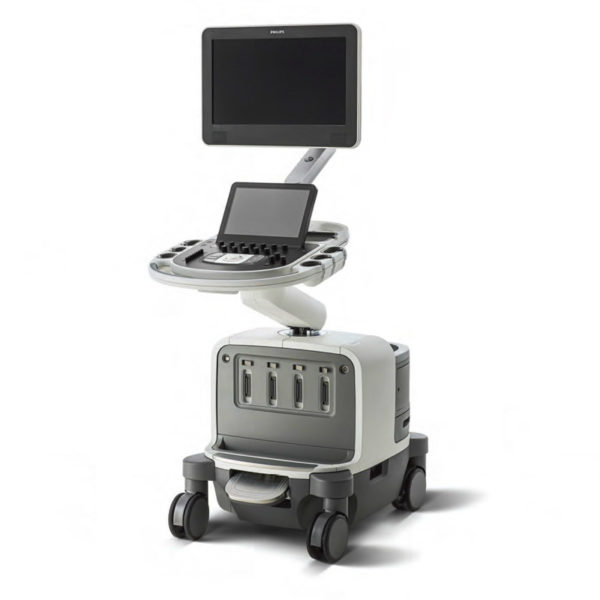
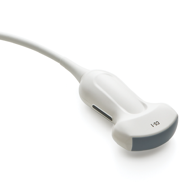
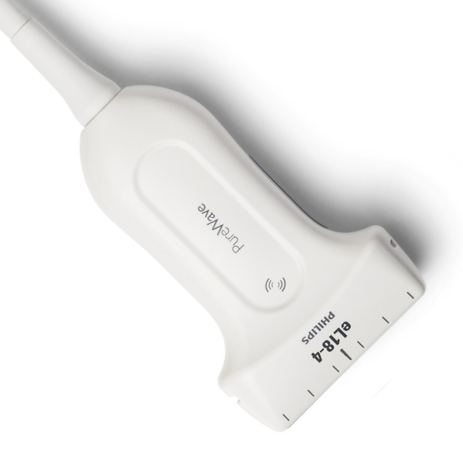
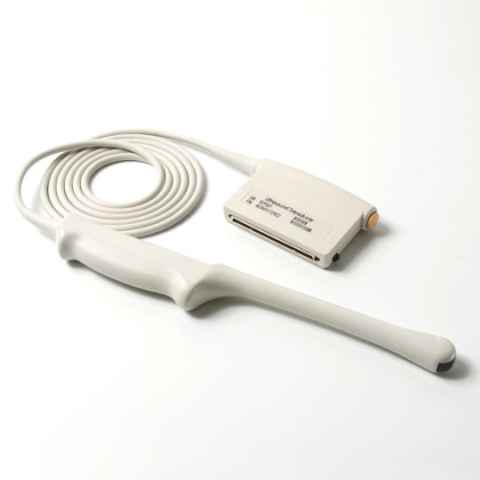
Ultrasound medical is a combination of acoustics, medicine, optics and electronics. Any application of acoustic technology above audible sound frequencies in the medical field is ultrasound medical. Including ultrasound diagnostics, ultrasound therapy and biomedical ultrasound engineering, so ultrasound medical has the characteristics of combining medicine, science and engineering, involving a wide range of content, and has high value in the prevention, diagnosis and treatment of diseases.
Ultrasound imaging scans the human body with ultrasonic sound beams, and obtains images of internal organs by receiving and processing reflected signals. There are many kinds of commonly used ultrasonic instruments: Type A (amplitude modulation type) indicates the strength of the reflected signal by the amplitude of the wave, and it shows an "echo graph". The M type (spot scanning type) represents the spatial position from shallow to deep in the vertical direction, and the time in the horizontal direction, which is displayed as the movement curve of the light spot at different times. The above two types are one-dimensional display, and the scope of application is limited. Type B (brightness modulation type) is an ultrasonic section imager, referred to as "B-ultrasound". The intensity of the received signal is represented by light spots with different brightness. When the probe moves along the horizontal position, the light spots on the display screen also move synchronously in the horizontal direction, and the trajectory of the light spots is connected to form a section view scanned by the ultrasonic sound beam. for two-dimensional imaging. As for the D-type is made according to the principle of ultrasonic Doppler. Type C uses a scanning method similar to that of a TV to display a cross-sectional acoustic image that is perpendicular to the sound beam. In recent years, ultrasonic imaging technology has been continuously developed, such as gray-scale display and color display, real-time imaging, ultrasonic holography, penetrating ultrasonic imaging, ultrasonic tomography, three-dimensional imaging, intracavity ultrasonic imaging, etc.
Ultrasound imaging are often used to determine the location, size, and shape of organs, determine the scope and physical properties of lesions, provide anatomical maps of some glandular tissues, and identify normal and abnormal fetuses. Digestive system, urinary system is widely used.

 English
 Русский


