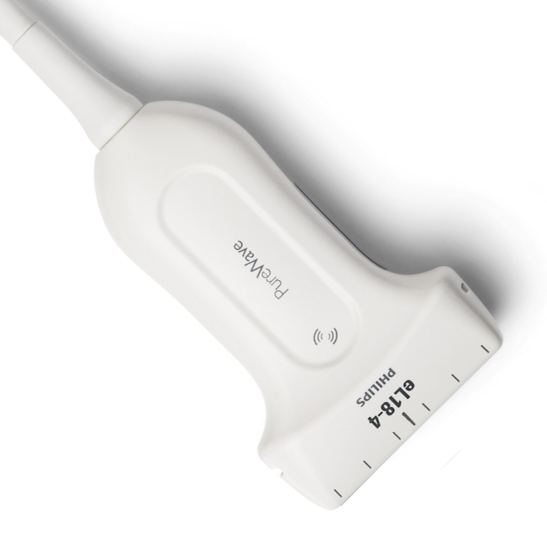
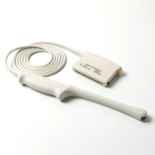
Ultrasound probes are crucial in diagnostic imaging processes in current medical technology. These little portable gadgets are an essential component of ultrasound scanners, allowing doctors to peek into the human body without intrusive treatments. Keep reading this article, and learn more about ultrasound probes.
The Basics Of Ultrasound Imaging
Ultrasound imaging, also known as sonography, is a widely used non-invasive medical imaging technique that utilizes high-frequency sound waves to produce real-time images of internal structures within the body. Unlike X-rays and CT scans that use ionizing radiation, ultrasound imaging is considered safer as it relies on harmless sound waves.
The ultrasonic probe, also known as a transducer, is the first step in the ultrasound imaging process. When pressed on the skin, this portable gadget releases sound waves into the body. After contacting materials of differing densities, these sound waves pass through different tissues and bounce back to the probe. The returning sound waves are then converted into electrical signals, which are processed by a computer to generate detailed images on a monitor.
What Is An Ultrasound Probe?
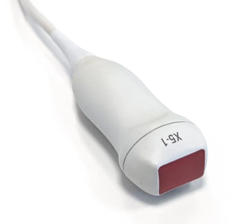
At the core of ultrasound imaging lies the ultrasound probe, also known as a transducer. This essential device is the workhorse that facilitates the entire imaging process. Ultrasound probes are typically small, handheld devices that come in various shapes and sizes, each designed for specific imaging purposes.
The key component of an ultrasound probe is the piezoelectric crystals it contains. These crystals are remarkable in that they can transform electrical energy into sound waves and sound waves back into electrical signals.
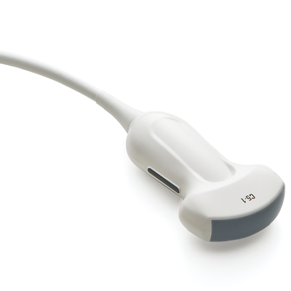
The design of the ultrasound probe is critical in determining the quality and resolution of the resulting images. Arrays of piezoelectric crystals set in various configurations on different probes govern the direction and focus of the generated sound waves. On the other hand, curvilinear probes have crystals arranged in a curved shape, providing broader images suitable for abdominal and obstetric imaging.
How Do Ultrasound Probes Work?
The working principle of ultrasound probes is based on the piezoelectric effect exhibited by the crystals they contain. When an electrical voltage is applied to these crystals, they vibrate and generate high-frequency sound waves. These sound waves travel into the body and interact with the tissues encountered along their path.
When the sound waves encounter a boundary between two tissues of different densities, such as the interface between muscle and bone, a portion of the sound waves is reflected back to the probe. The probe's crystals then switch to the receive mode, converting the returning sound waves into electrical signals. These signals are transferred to the ultrasound machine, which processes them and converts them into visual representations on the display.
By measuring the time it takes for the sound waves to travel to and from different structures within the body, the ultrasound machine creates precise spatial information. This information is then utilized to create real-time pictures, allowing healthcare providers to see organs, blood arteries, and other inside structures in incredible depth and precision.

Conclusion
The mobility, safety, and adaptability of ultrasound probes have made them important in a variety of medical disciplines. If you are looking for a new ultrasound probe in the market, look no further than Xity! Browse our website for more product details now!
 English
 Русский

