English
 Русский

In our time, 3D / 4D volumetric ultrasound has become very widespread, especially in the field of obstetrics. On the facades of private, and often budgetary, medical institutions, on the Internet, in the media, you can often find an advertisement for such a service with beautiful pictures and promises of an unforgettable experience recorded on a CD or other media. What is behind all these?
Let's start with the terms again. Routine ultrasound is performed in 2D mode (D, dimension - measurement; synonymous with B-mode), when two-dimensional "flat" gray-scale ultrasound slices are obtained, which are very updated at a speed of more than 20 frames per second, which is perceived by the human eye as real-time video. Additionally, the so-called Doppler modes (color and spectral) are used, which allow obtaining qualitative and quantitative information about the blood flow in the studied vessels.
In 3D mode, a third spatial dimension appears - depth. A 3D examination captures multiple 2D slices, from which the corresponding software very quickly reconstructs one still 3D image, which can then be presented in a variety of ways. In 4D mode, 3D images are updated very quickly, the 3D image is perceived as a real-time movie.
Time is considered as another dimension (3+1), but the essence of the mode is similar to 3D. In order to ensure fast updating of the volumetric image over time, the system captures 2D images at a lower density, so the quality (“density”) of volumetric information in 4D mode is lower than in 3D mode. Specialists are often forced to use only the 4D mode if the object under study (most often a fetus in the uterus) is actively moving. 3D/4D is always an additional time investment.
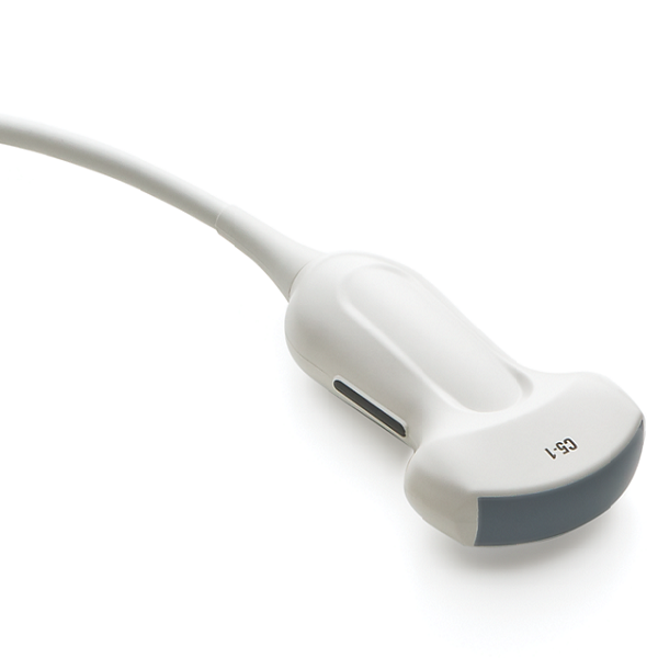
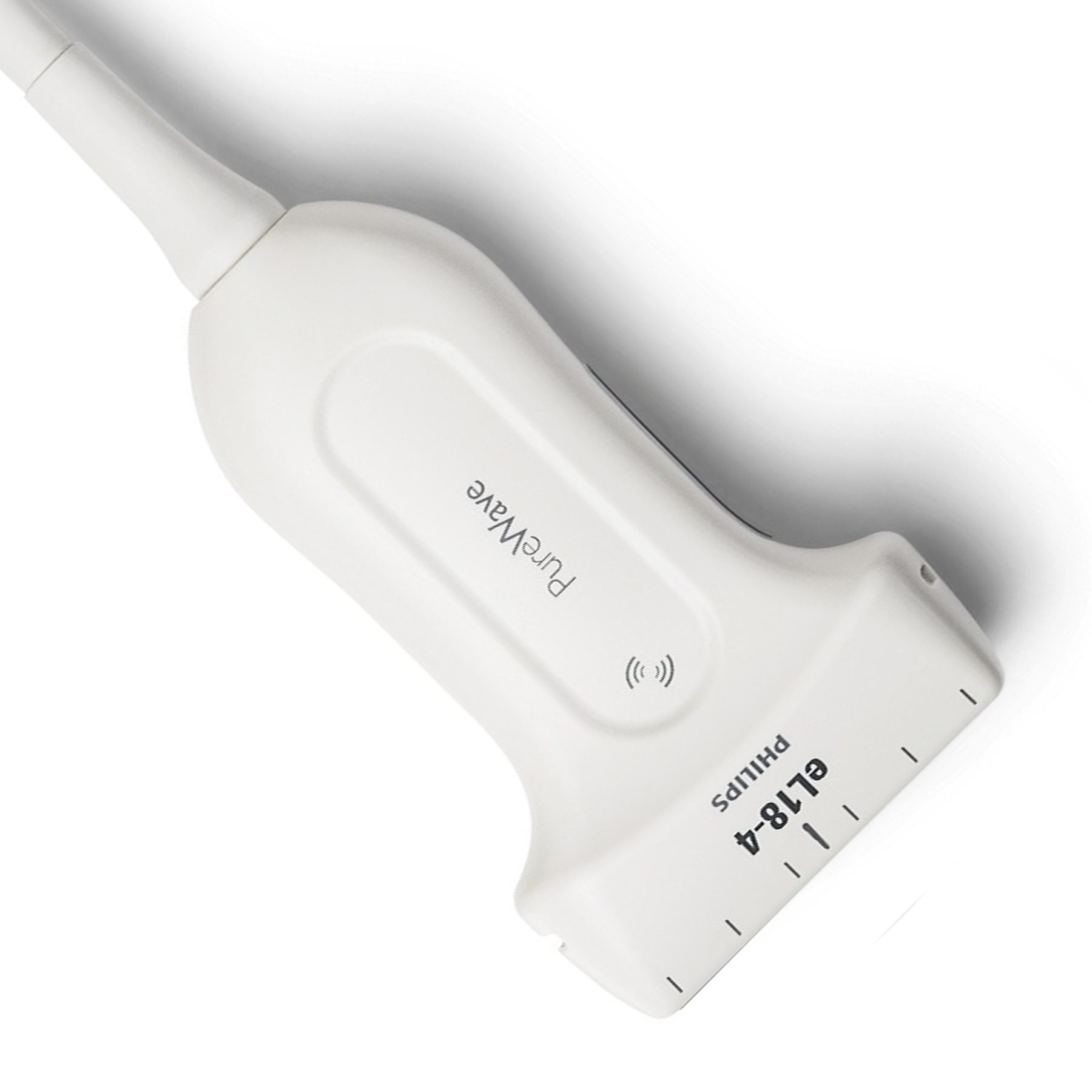
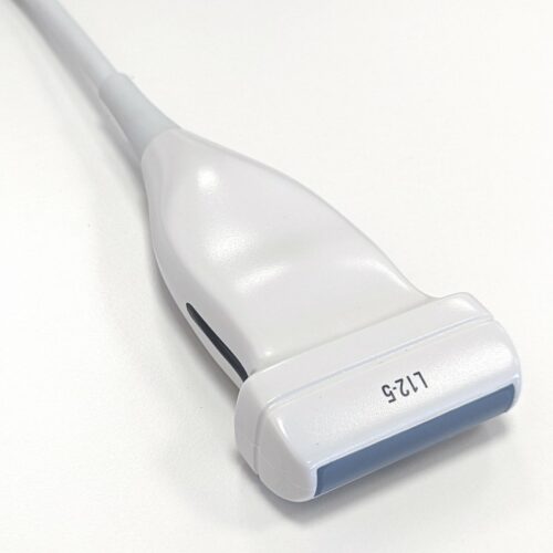
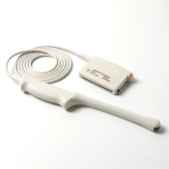
To obtain 3D/4D images, special volumetric sensors are used, as a rule they are larger and heavier than sensors for routine studies. During the examination, the patient may feel a slight vibration from such sensors, because. there are moving components inside when the sensor is operating in volumetric mode. Progress does not stand still, the latest models of volumetric sensors (especially the so-called "matrix") almost do not differ in appearance and dimensions from conventional sensors.
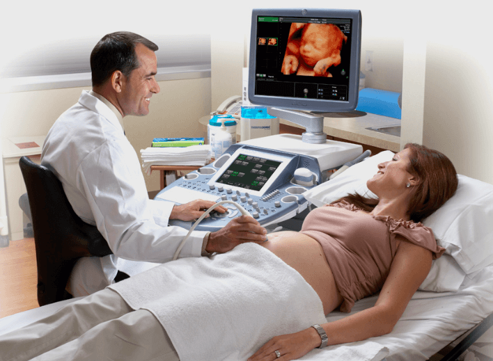
An ultrasound machine looks like a conventional one; special software and specialized sensors are required to obtain a three-dimensional image. In part, the class of the device can be judged by the size of the monitor (on expert ones from 19 inches or more) and the presence of a touch panel (“touch-screen”) on the control panel of the device.
We are used to the fact that 3D/4D ultrasound is an ultrasound that is usually used to take a photo or video of the fetal face, legs, arms, genitals, etc. This is the so-called surface volumetric reconstruction mode (photorealistic, surface, etc.). At short stages of pregnancy (up to about 13-14 weeks), you can get a surface volumetric image of the entire fetus.
The most realistic facial image can be obtained at 22-30 weeks, when human features are already more formed and there are conditions for obtaining a high-quality surface reconstruction. On the best modern instruments, there are a variety of options for this type of three-dimensional image, sometimes even “frighteningly realistic”.
A number of studies have been conducted on the effects of 3D ultrasound technologies on the human fetus: no harmful effects of 3D / 4D ultrasound have been identified. A wave of publications against the use of 3D / 4D ultrasound, which appeared at one time in the media, was aimed at non-medical, non-diagnostic use of three-dimensional ultrasound, when ultrasound devices began to be purchased privately, installed almost in hairdressing salons and many hours (!!) of monitoring the behavior of the fetus was carried out in the womb in 4D.
It is usually used to show a woman her baby in motion. For diagnostic purposes, static 3D modes are mainly used.