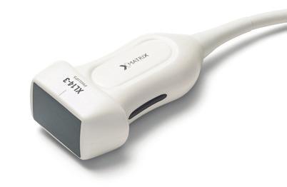
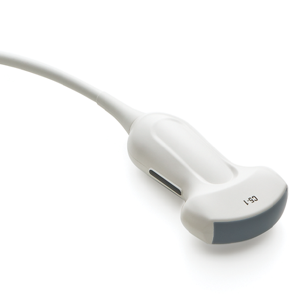
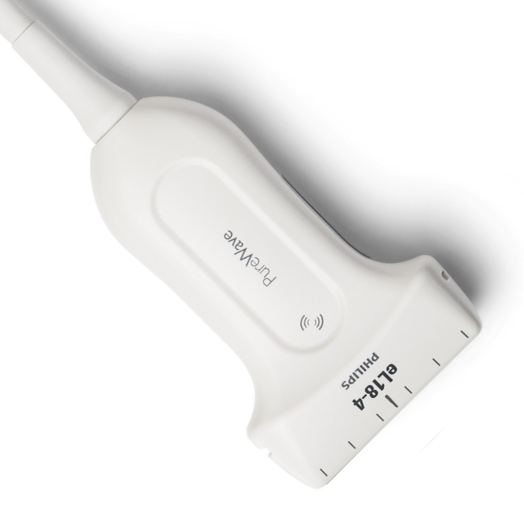
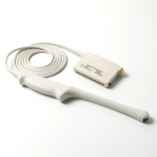
Ultrasound technology has transformed medical imaging by providing non-invasive, real-time viewing of interior structures. Ultrasound probes, both two-dimensional (2D) and three-dimensional (3D), are widely employed in obstetrics, gynecology, cardiology, and other medical disciplines. While both serve the same fundamental purpose of capturing images inside the body, they differ significantly in their capabilities and applications. In this article, we will delve into the dissimilarities between 2D and 3D ultrasound probes, shedding light on the unique advantages each technology brings to the world of medical diagnostics.
Image Representation
2D Ultrasound
2D ultrasound, often referred to as conventional ultrasound, relies on beams of sound waves that bounce off tissues and organs, creating a two-dimensional image on the ultrasound monitor. Because this technology is capable of recording dynamic, real-time pictures, it is ideal for monitoring moving structures such as a growing baby during pregnancy. The simplicity of the images aids in quick interpretation and is widely used for routine examinations.
3D Ultrasound
In contrast, 3D ultrasound utilizes advanced technology to capture multiple 2D slices from various angles, creating a volumetric representation of the scanned area. This results in more realistic and detailed pictures, providing for a more thorough view of organ surfaces and contours. The added dimension improves diagnostic skills, allowing for a more detailed knowledge of anatomical structures.

Visualization And Detail
2D Ultrasound
The strength of 2D ultrasound lies in its ability to visualize movement and real-time processes. As a result, healthcare providers may watch the baby's movements, heartbeat, and growth, making it a great tool for tracking fetal development. However, the lack of depth perception may limit the detailed visualization of complex anatomical structures.
3D Ultrasound
3D ultrasound excels in providing detailed anatomical information, offering a more realistic and immersive visualization of internal structures. It enables healthcare personnel to more clearly analyze the surfaces of organs and structures, allowing for a more in-depth investigation of complicated forms. This capability is particularly beneficial in situations where a more detailed view is essential for accurate diagnosis and treatment planning.

Applications
2D Ultrasound
2D ultrasound is the go-to choice for routine examinations during pregnancy, offering real-time insights into fetal development. Furthermore, it is commonly used for abdominal, pelvic, and cardiac imaging, where real-time monitoring is critical for diagnosis and patient management.
3D Ultrasound
3D ultrasound finds a market in applications that require more comprehensive anatomical viewing. It is frequently used for extensive prenatal exams, enabling for a thorough study of the growing fetus. Furthermore, it is valuable in cardiac imaging, where the intricate structures of the heart benefit from the enhanced spatial information provided by 3D technology.

Diagnostic Accuracy
2D Ultrasound
While 2D ultrasound is a viable diagnostic tool, particularly for real-time monitoring and routine exams, its diagnostic accuracy may be restricted when dealing with complex anatomical structures or minor anomalies. The interpretation heavily relies on the expertise of the healthcare professional.
3D Ultrasound
3D ultrasound contributes to heightened diagnostic accuracy by offering a more comprehensive view of the scanned area. The additional dimension and increased detail enhance the ability to detect anomalies and abnormalities that might be challenging to identify using 2D imaging alone. This can lead to more accurate diagnoses and better patient outcomes.
Conclusion
The decision between 2D and 3D ultrasound probes in medical imaging is determined by the unique diagnostic criteria and type of medical examination. Both technologies play crucial roles in enhancing medical professionals' ability to visualize and understand the intricacies of the human body, contributing to improved patient care and diagnostic accuracy. Besides, if you are looking for a suitable ultrasound probe for your hospital, Xity is ideal for you. Contact us for more details now!
FAQs
1. Which ultrasound probe is better for routine pregnancy monitoring, 2D, or 3D?
Answer: While both 2D and 3D ultrasound probes serve essential roles in pregnancy monitoring, 2D ultrasound is typically the preferred choice for routine examinations during pregnancy. Its real-time imaging capabilities are well-suited for tracking fetal development, observing movements, and monitoring the baby's heartbeat. 3D ultrasound, on the other hand, is often utilized for more detailed assessments, providing a comprehensive view of the fetus's features and surfaces.
2. Are there any additional safety concerns with 3D ultrasound compared to 2D?
Answer: Both 2D and 3D ultrasound technologies are considered safe for medical imaging purposes. The sound waves used in ultrasound scans are non-ionizing and have no known harmful effects on the human body. However, it's important to note that 3D ultrasound may require longer scanning times and may involve higher energy levels, but these factors are well within established safety guidelines. As always, ultrasound procedures should be performed by trained healthcare professionals following recommended safety protocols.
3. Can 3D ultrasound replace 2D ultrasound in all medical applications?
Answer: While 3D ultrasound offers enhanced visualization and is particularly valuable in certain applications, it may not completely replace 2D ultrasound in all medical scenarios. 2D ultrasound remains a versatile and effective tool, especially in real-time monitoring and routine examinations. The choice between 2D and 3D ultrasound depends on the specific diagnostic requirements of each medical situation. In some cases, a combination of both technologies may be employed to provide a comprehensive assessment of the patient's condition.
 English
 Русский


