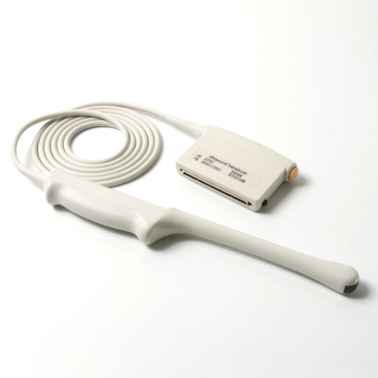
Ultrasound technology has proven to be a vital component in medical diagnostics and an array of other applications. Ultrasound probes are differentiated primarily by their frequency, with low-frequency (2-5 MHz) and high-frequency (7-18 MHz and above) variants serving as the primary categories. Grasping the nuances between high-frequency and low-frequency ultrasound probes is essential for their effective utilization in medical imaging, industrial testing, and beyond. This exploration will guide you through the fundamental distinctions, practical applications, and unique benefits of varying ultrasound probe frequencies, ensuring a comprehensive understanding for healthcare professionals and technicians alike.
Low-Frequency Ultrasound Probes
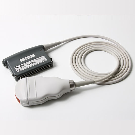
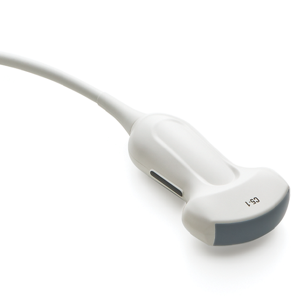
The realm of low-frequency ultrasound probes is characterized by their ability to penetrate deep into the body, making them ideal for visualizing larger, deeper structures. Operating typically between 2-5 MHz, these probes can traverse through dense tissue, reaching organs such as the liver and kidneys, and are invaluable in obstetric imaging for assessing fetal development. However, the trade-off for this deep penetration is a lower imaging resolution, resulting in images that may appear grainier and less defined. Despite this, their proficiency in scanning extensive areas makes them indispensable in evaluating the overall condition and integrity of internal structures. Clinically, low-frequency probes are pivotal in specialties like obstetrics, gynecology, and abdominal imaging, where a comprehensive view takes precedence over fine detail.
Penetration Depth: Low-frequency ultrasound probes, with their 2-5 MHz frequency range, are adept at deep-tissue imaging. This lower frequency spectrum allows the ultrasound waves to penetrate further, making them capable of reaching significant depths within the body, such as visualizing the liver, kidneys, or fetus during prenatal care.
Imaging Resolution: While low-frequency ultrasound probes offer extensive penetration, this comes at the cost of imaging clarity. The lower frequencies are not as adept at delineating fine details, which can result in somewhat coarser images, with broader structure outlines. Nonetheless, these probes are highly effective for surveying expansive areas and evaluating the general health and robustness of deep-lying anatomical features.
Clinical Applications: Low-frequency ultrasound probes are widely used in a range of therapeutic settings, including obstetrics and gynecology for fetal monitoring and abdominal organ evaluation. They are particularly valuable for imaging larger anatomical structures where the clinical priority is to ensure proper function and detect abnormalities at a macroscopic level. They are also widely employed in musculoskeletal and vascular imaging.
The capacity of low-frequency probes to penetrate deeply and study huge regions is their principal benefit. In cases where assessing structural integrity and overall function is more critical than pinpointing small abnormalities, such as evaluating the size and shape of organs or checking for vascular blockages, low-frequency probes are indispensable.
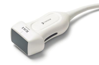
Philips XL14-3 XMatrix Ultrasound Probe
High-Frequency Ultrasound Probes
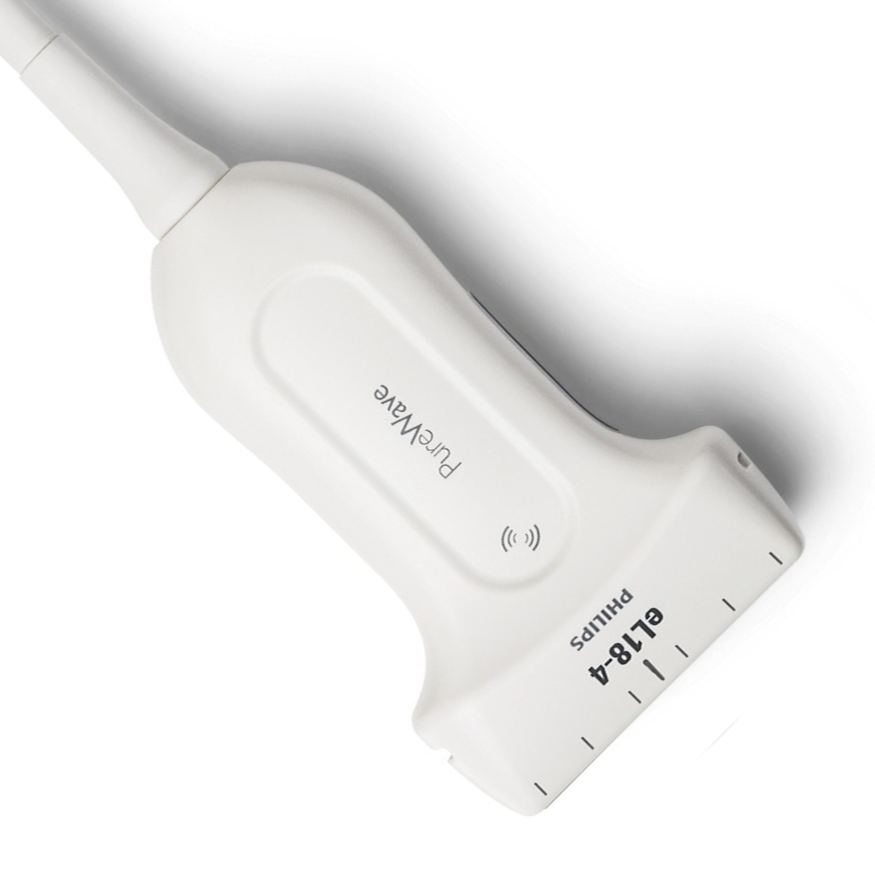
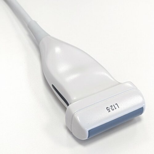
Transitioning to the sphere of high-frequency ultrasound probes, we encounter devices designed for precision. These probes, operating within the 7-18 MHz range or higher, are tailored for superficial examinations, offering high-resolution images that are crucial for detecting minute details. Although their penetration is limited compared to their low-frequency counterparts, the level of detail they can provide is unparalleled. This makes them particularly useful for specialties such as dermatology, where skin layers can be examined in detail, and ophthalmology, where the fine structures of the eye require meticulous observation.
Here are some key features of High-Frequency Ultrasound Probes:
Penetration Depth: High-frequency ultrasound probes typically operate in the range of 7-18 MHz or even higher. These frequencies result in shallower penetration but offer exceptional resolution for fine details. High-frequency ultrasound is limited to superficial structures, as the waves do not penetrate deeply into the body.
Imaging Resolution: The capacity of high-frequency probes to provide high-resolution pictures with exceptional clarity is its distinguishing attribute. These probes excel at capturing fine details and differentiating between subtle abnormalities. The images are characterized by sharp, detailed contours, making them ideal for early disease detection and precise measurements.
Clinical Applications: High-frequency ultrasound probes have a wide array of applications across diverse medical specialties. Dermatologists employ these probes for detailed imaging of skin lesions and for assessing the extent of skin cancers. In ophthalmology, the probes are indispensable for conducting precise evaluations of the cornea and retina. Additionally, they are crucial in breast imaging and the examination of small parts, where high-resolution imaging is necessary to distinguish between benign and malignant lesions.
The principal advantage of high-frequency probes lies in their ability to produce detailed and sharp images of superficial structures. This attribute is invaluable for early diagnosis and precise intervention in medical fields where the discernment of fine anatomical details is crucial.
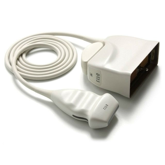
Philips L17-5 High-frequency Linear Probe
Selecting the Appropriate Ultrasound Probe
The decision-making process for selecting the right ultrasound probe is nuanced and depends on the specific requirements of the clinical or industrial application at hand. In the medical context, choosing between low and high-frequency probes hinges on the targeted anatomical depth and the desired level of detail in the images. Low-frequency probes are the go-to option for imaging deep-seated body structures, whereas high-frequency probes are preferred for detailed surface analysis. In industrial settings, the nature of the material under inspection and the depth of potential defects are critical factors influencing probe selection. High-frequency probes are typically chosen for examining thin materials, whereas low-frequency probes are favored for thicker materials where deep penetration is necessary to identify potential faults.
Conclusion
The key difference between low and high-frequency ultrasound probes lies in their penetration depth and imaging resolution. Choosing the right probe depends on the specific application's requirements. If you are looking for an ultrasound probe for your business, Xity is ideal for you. With more than 10 years experience of in offering nationwide repair, service, maintenance, distribution, and sales of diagnostic ultrasound systems, probes, and parts. Choose us and get high-quality products!
 English
 Русский

