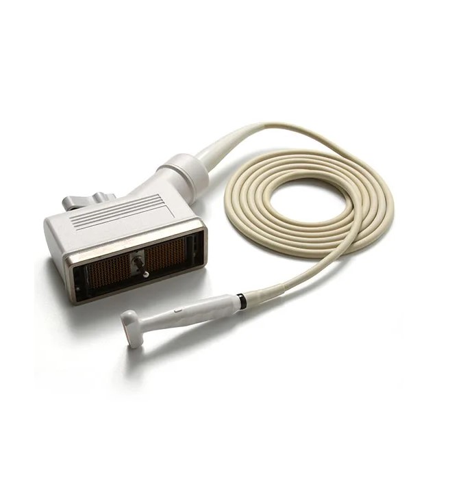
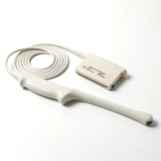
Ultrasound technology has significantly advanced medical diagnostics, providing healthcare professionals with a non-invasive method to visualize the body's internal structures with high accuracy and safety. Among the various tools at their disposal, linear and convex ultrasound probes stand out due to their distinct characteristics and specialized applications. This article aims to explore the fundamental differences between linear and convex ultrasound probes, enhancing understanding of their optimal uses in clinical practice.
Linear Ultrasound Probes
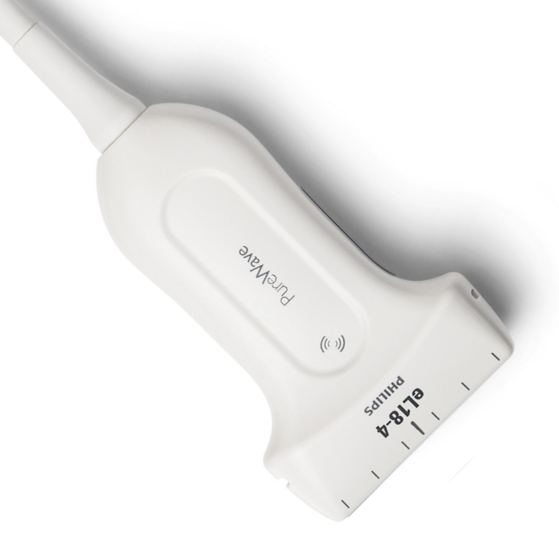
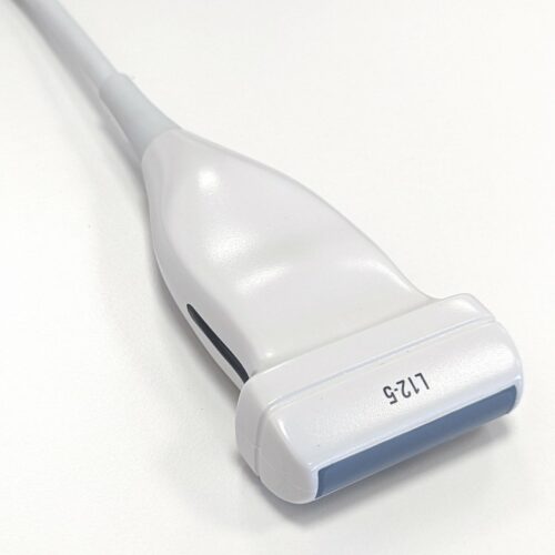
Linear ultrasound probes are distinguished by their straight-edged, rectangular transducers that linearly emit sound waves. These probes are specifically designed for high-frequency imaging, which is optimal for detailed visualization of superficial structures that lie just beneath the body's surface. Here are some notable features and applications of linear ultrasound probes:
High-Frequency Imaging: Linear probes operate at frequencies ranging higher. Because of the high frequency, they provide excellent picture quality and detail, making them ideal for superficial exams. Linear probes work at higher frequencies.
Musculoskeletal Imaging: Linear probes are commonly used for musculoskeletal examinations, including the assessment of tendons, ligaments, muscles, and joints. They are especially helpful in orthopedics for assessing injuries and directing therapies such as injections.
Vascular Imaging: These probes are also employed for vascular studies, especially for assessing blood vessels close to the skin's surface. They can identify irregularities in blood flow, such as deep vein thrombosis (DVT) or varicose veins.
Soft Tissue Imaging: Linear probes excel at visualizing soft tissues, including the thyroid, breast, and superficial masses. They're useful for identifying malignancies and tracking their development.
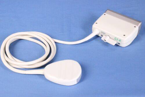
Convex Ultrasound Probes
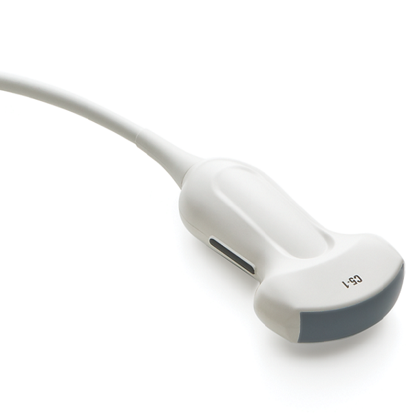
Alternatively, convex ultrasound probes are equipped with a curved transducer surface that projects sound waves in a divergent, fan-shaped pattern. These probes operate at lower frequencies, which is advantageous for penetrating deeper into the body and inspecting internal regions that are not as accessible with high-frequency probes.
Here are some key characteristics and applications of convex ultrasound probes:
Lower Frequency Imaging: Convex probes typically operate at frequencies ranging from 2 to 7 MHz. The lower frequency allows for more penetration and is ideal for imaging organs deeper within the body.
Abdominal Imaging: Convex probes are widely used for abdominal scans, including the liver, spleen, kidneys, and uterus. They have a wider field of vision and are ideal for assessing the size and function of these organs.
Obstetrics and Gynecology: Convex probes are preferred for prenatal imaging in obstetrics because they can acquire detailed pictures of the growing baby. They are also used in gynecology to assess the uterus and ovaries.
Cardiac Imaging: Convex probes can be used for echocardiography to see the chambers and valves of the heart. Their wide field of view is beneficial for cardiac assessments.

Distinguishing Between Them
It is vital for healthcare practitioners to distinguish between linear and convex ultrasonic probes in order to make educated judgments when selecting the proper probe for a certain medical imaging task. Here are the differences between them:
Frequency
Linear ultrasound probes operate at higher frequencies, typically in the range of 7 to 15 MHz or even higher. This high frequency allows for exceptional image resolution and is well-suited for imaging superficial structures and tissues. Convex ultrasonic probes, on the other hand, operate at lower frequencies, often ranging from 2 to 7 MHz. Because of the lower frequency, they can penetrate deeper into the body, making them suitable for imaging deeper tissues and bigger organs.
Shape
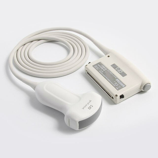
Linear probes have a flat or rectangular transducer surface. This flat shape emits sound waves in a straight line, which is suitable for capturing high-resolution images of structures close to the skin's surface. The transducer surface of convex probes is curved. This curved structure generates sound waves in the form of a fan-shaped beam, providing for a larger field of vision and deeper imaging capabilities.
Applications
Linear ultrasound probes are best suited for applications that require high-resolution imaging of superficial structures. When examining bigger, deeper organs and tissues, convex ultrasonic probes are frequently employed.
Field Of View
Due to their high-frequency operation and narrow beam, linear probes provide a relatively limited field of view. They excel at capturing fine details in a small area. Convex probes have a greater field of view and are thus suited for imaging bigger anatomical areas. When analyzing the size, shape, and general condition of organs or structures located deeper within the body, this larger view is useful.
Conclusion
Linear and convex ultrasound probes are integral to the modern landscape of medical imaging, each offering distinct advantages for various diagnostic applications. Xity, renowned as one of the famous ultrasound equipment suppliers in China, offers an extensive range of not only ultrasound probes but also essential ultrasound parts. For those interested in expanding their diagnostic capabilities, we invite you to contact us for more comprehensive product information and details today!
FAQs
1. What are the main uses of linear ultrasound probes?
Linear ultrasound probes are primarily used for high-resolution imaging of superficial body structures. Their high-frequency sound waves make them ideal for detailed assessments in musculoskeletal imaging, which includes examining muscles, tendons, ligaments, and joints. They are also widely used for vascular imaging to detect conditions like deep vein thrombosis, as well as for evaluating soft tissues such as the thyroid, breast, and superficial masses.
2. When would I choose a convex ultrasound probe over a linear one?
Convex ultrasound probes are chosen for their ability to image deeper structures within the body, thanks to their lower-frequency sound waves. They are particularly effective for abdominal imaging, providing a broad field of view necessary for evaluating organs like the liver, spleen, and kidneys. Additionally, convex probes are the preferred choice in obstetric and gynecological imaging for their capacity to capture detailed images of the fetus during pregnancy and assess the uterus and ovaries.
3. How does the frequency of an ultrasound probe affect imaging?
The frequency of an ultrasound probe is directly related to its imaging capabilities. Higher-frequency probes, such as linear probes, offer high resolution and are excellent for visualizing fine details in superficial tissues. However, their penetration depth is limited. Lower-frequency probes, like convex probes, have greater penetration, allowing for visualization of deeper tissues and organs but with slightly reduced image resolution. The choice between high and low-frequency probes depends on the specific clinical requirements of the examination.
 English
 Русский

