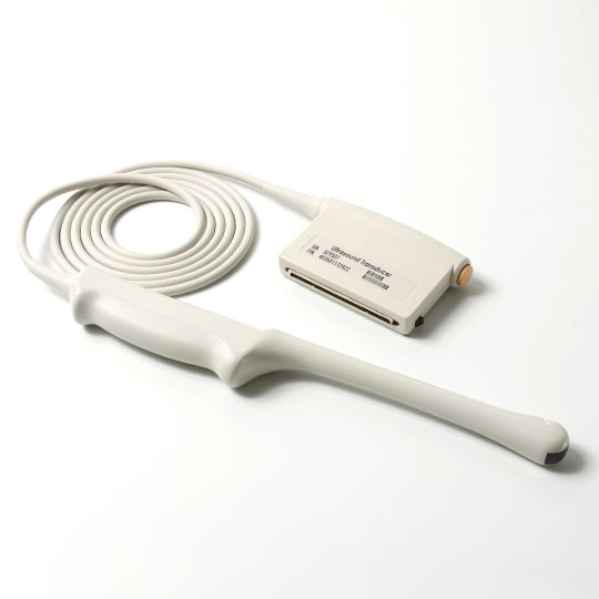
The advent of Transesophageal Echocardiography (TEE) has been a game-changer in the realm of cardiac care, offering unparalleled insights into the heart's structure and function. This article delves into the intricacies of the TEE ultrasound probe, a pivotal instrument in modern cardiology that enhances diagnostic precision and patient care.
Introduction to TEE Ultrasound Probes
Transesophageal Echocardiography (TEE) represents a significant leap forward in cardiac imaging. By utilizing a specialized probe, called a TEE probe or transducer, cardiologists can obtain highly detailed images of the heart from an esophageal vantage point. This method circumvents the common obstacles encountered with transthoracic echocardiography, such as lung air or ribcage interference, thereby providing a clearer window into cardiac anatomy and pathology.
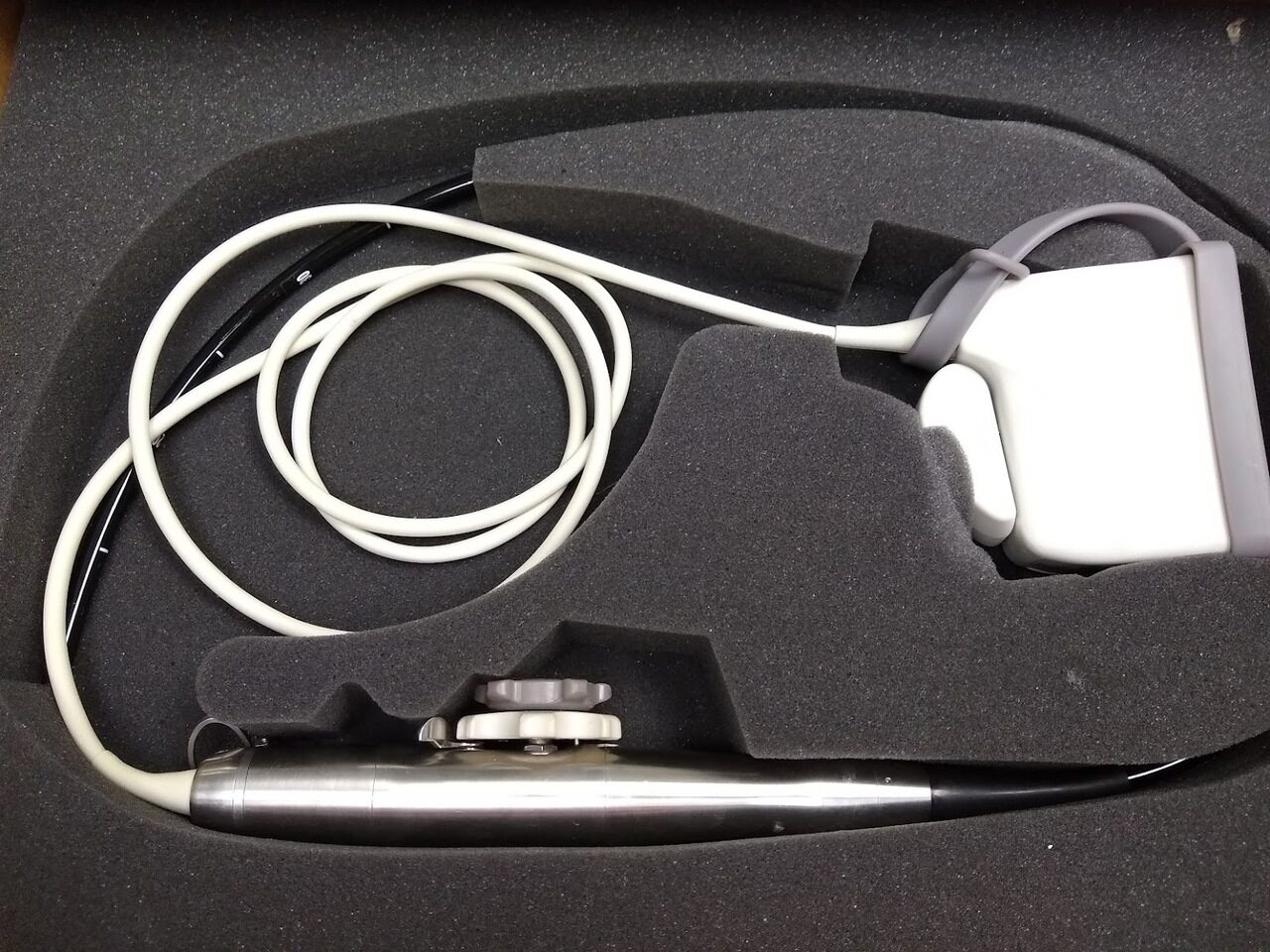
The TEE Ultrasound Probe
A TEE probe, also known as a TEE transducer, is an advanced medical device designed for use in TEE diagnostic procedures. It comprises a flexible shaft equipped with an ultrasound transducer and a camera at its tip, which are inserted into the esophagus after appropriate anesthesia is administered to the patient. The proximity of the esophagus to the heart allows the TEE probe to generate high-resolution images that are critical for accurate diagnosis and management of cardiac diseases.
How Does A TEE Ultrasound Probe Work?
The TEE ultrasound probe functions similarly to regular ultrasound technology. It sends high-frequency sound waves (ultrasound) into the body, which bounce off interior structures and cause echoes. By analyzing the echoes that return to the probe, the system generates real-time images of the heart and nearby vessels.
The TEE probe's position in the esophagus allows it to capture exceptionally clear and detailed images of the heart's chambers, valves, and blood flow. This closeness is especially useful for detecting diseases such as valve anomalies, blood clots, congenital heart defects, or structural concerns that may be difficult to detect with regular echocardiography.
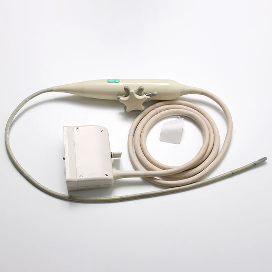
The Benefits Of TEE Ultrasound Probe
Let's delve deeper into the benefits of the TEE ultrasound probe:
Enhanced Image Quality
One of the standout advantages of TEE is the exceptional image quality it provides. Positioning the ultrasound probe close to the heart overcomes many of the limitations associated with traditional echocardiography, such as interference from air in the lungs or the ribcage. TEE provides crisper, higher-resolution pictures of the heart's components as a consequence, making it simpler to spot minor anomalies and deliver more accurate diagnoses.
Real-Time Imaging
TEE provides real-time imaging of the heart, allowing healthcare providers to assess cardiac function dynamically. This capacity is especially useful during surgical procedures since it allows physicians to monitor the heart's function and adjust their activities accordingly.
Improved Diagnosis
The TEE ultrasound probe is an invaluable tool for diagnosing a wide range of cardiac conditions. It excels at detecting structural abnormalities, valve dysfunction, and congenital heart defects that may be challenging to identify through other imaging modalities. The accuracy and precision of TEE allow for earlier diagnosis and intervention, resulting in more successful treatment approaches and improved patient outcomes.
Reduced Invasiveness
Compared to other diagnostic techniques that may require invasive procedures, such as cardiac catheterization, TEE is minimally invasive. To alleviate the pain of probe insertion, patients undergoing TEE often receive conscious sedation and local anesthetic. This decreases the need for general anesthesia and the hazards connected with it.
Monitoring Critical Care Patients
In critical care settings, TEE is a powerful tool for continuously monitoring patients with acute cardiac conditions. It gives healthcare personnel rapid insight into the heart's function, allowing them to make quick changes to treatment strategies. For patients in severe conditions, real-time monitoring can be lifesaving.
Conclusion
The Impact of TEE Ultrasound Probes The TEE ultrasound probe has profoundly impacted cardiology, offering a minimally invasive yet highly accurate window into the heart's functioning. Its contribution to the early detection and treatment of cardiac conditions underscores its indispensable role in contemporary medical practice.
For healthcare providers seeking top-notch cardiac imaging solutions, Xity stands out as a premier supplier of ultrasound probes, including state-of-the-art TEE ultrasound probes. Trust Xity for your ultrasound needs and elevate your patient care to new heights.
 English
 Русский

